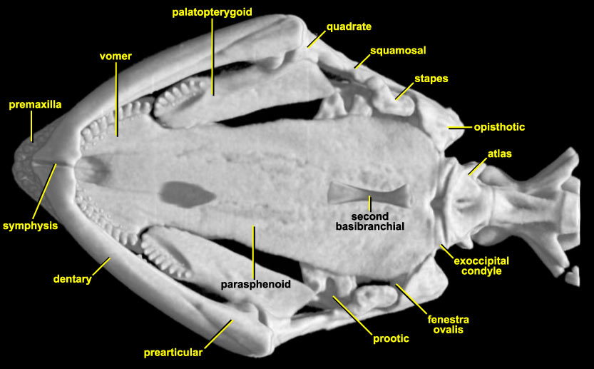
In the absence of maxillae, the premaxillae and vomers dominate the anterolateral part of the skull, with the premaxillary and vomerine teeth forming parallel arches. Posterolaterally, the vomers meet narrow plate-like palatopterygoids. These also bear a short marginal tooth row, leading some authors (e.g., Trueb, 1993) to suggest that this bone may be a compound of pterygoid and vestigial palatine. Further posteriorly, the pterygoid abuts the medial surface of the quadrate. In the midline, the remainder of the palate is provided by the parasphenoid. This bone is narrow anteriorly, extending between the vomers almost to the tip of the skull. Behind the vomers, the parasphenoid broadens out, reaching its greatest width just behind the quadrates. Trueb (1993) notes that the parasphenoid wings are perforated by foramina for the internal carotid artery, but this does not appear to be the case in this specimen. The prootic and opisthotic are visible in ventral view, and are separate. The fenestra ovalis is filled by a large stapes with a distinct stylus. This contacts a process from the squamosal (use the Skeleton Only roll movie to see this more clearly). This view also shows clearly the relationship between the paired exoccipital condyles and the first vertebra, the atlas. Apparently floating below the palate is a slender, finger-like bone. This is the second basibranchial element of the hyoid. Most of the hyoid in Necturus is comprised of paired lateral arches, with median basibranchial elements. Only the second of these median elements is ossified, and thus visible in the CT imagery.
The lower jaw frames the palate in this view and shows clearly the anteromedial symphysis between the tooth-bearing dentaries. Two other bones make up the jaw of Necturus, and again the specimen needs to be rolled to see them clearly. Behind the dentary on the inferior surface, but extending forward along the medial surface, there is the triangular prearticular bone (incorporating the angular). Behind the dentary tooth row, there is a third, smaller bone, bearing a short tooth row that is continuous with that of the dentary. This bone is the coronoid (operculare, Wiedersheim, 1877; splenial, Hecht, 1957). The presence of the coronoid is unusual amongst living salamanders (see Dicamptodon for another example), as is the fact that it bears teeth. The posterior tip of Meckels cartilage, the articular, does not ossify so that the quadrate appears to meet an open space in the centre of the jaw.