Expert annotations for this species! See below. Expert annotations for this species! See below.
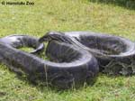
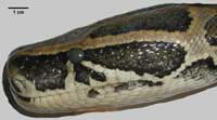
Python molurus is one of the worlds largest snakes, with records of up to 9.15 m in length. It belongs to the family Boiidae (subfamily Pythoninae) and is distributed across Asia from Pakistan to Indonesia and South China, with two distinct subspecies. P. m. molurus is the Indian python (Pakistan, India, Bangladesh, Nepal, Sri Lanka) while P. m. bivittatus is the Burmese python (Burma, Laos, Vietnam, Thailand, Peninsula Malaysia, Cambodia, Indonesia and South China). |
|
The Burmese python frequents forests near water bodies, swamps, marshes and grasslands. It can move easily in the trees, on the ground, and in water, and is a nocturnal hunter. It has special heat receptors to locate warm prey. Like other boiids, the Burmese python kills by constriction and suffocation, coiling around the prey and slowly tightening the coils further each time the victim breathes out. It does not crush. Prey animals include a wide range of taxa from small mammals to deer and even leopards.
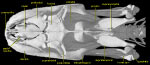
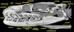
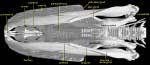
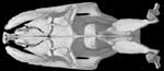
The skull of the Burmese python is very highly ossified, with dense bone and complex sutures. Like other snakes, it has lost the upper temporal bar, jugal, squamosal, and epipterygoid. Snakes have an inner ear and stapes, but no tympanum, no auditory meatus, and no tympanic cavity. A bony interorbital septum is present.
|
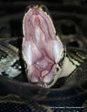
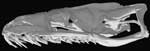
Python m. bivittatus is a macrostomatan (big-mouthed) snake that uses its exceptionally flexible (kinetic) skull to create an enormous gape. The upper and lower jaws, and the bones of the palate, are loosely attached to a rigid box comprising the braincase and part of the skull roof (frontals and parietal). The quadrates have a greater freedom of movement than in lizards, partly due to a reduced contact with the pterygoids. Thanks to the extended supratemporals, they are positioned well behind the craniocervical joint, further increasing the potential gape. Since the supratemporals are also loosely connected to the skull, they can swing out laterally, another factor affecting the size of the mouth and the width of the throat. There is no bony contact between the anterior ends of the dentaries (highly elastic ligament) so they are able to spread apart. |
As a result of these modifications, the python can swallow prey 4-5 times the diameter of its own head. The jaws and palatal elements can also move independently about the midline, and each bears a series of extremely sharp, posteriorly recurved teeth. These are used to draw the prey into the mouth using a ratchet action that alternates between the two sides of the skull.
Additional Information on the Skull
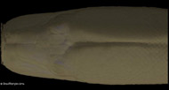
Click on the thumbnails below for a detailed description of the skull in standard anatomical views.
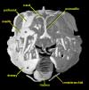
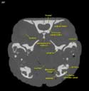
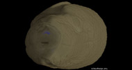
Click on the thumbnail below for a description of internal features of the skull based on selected coronal slices.

About the Species
About this Specimen
This specimen was scanned by Matthew Colbert on 9 July 2003 along the coronal axis for a total of 810 slices. Each 1024x1024 pixel slice is 0.1055 mm thick, with an interslice spacing of 0.1055 mm and a field of reconstruction of 45.0 mm.
About the
Scan
Literature
Baird, I. I. 1970. The anatomy of the reptilian ear, pp. 193-275. In Gans, C. and Parsons, T. S. (eds), Biology of the Reptilia, vol. 2, Academic Press, London and New York.
Frazzetta, T. H. 1959. Studies on the morphology and function of the skull in the Boidae (Serpentes). Pt 1: Cranial differences between Python sebae and Epicrates cenchris. Bulletin of the Museum of Comparative Zoology 119:453-472.
Gans, C. 1961. The feeding mechanism of snakes and its possible evolution. American Zoologist 1:217-227.
Kluge, A. G. 1991. Boine snake phylogeny and research cycles. Miscellaneous Publications of the Museum of Zoology, University of Michigan 178:1-58.
Kluge, A. G. 1993. Aspidites and the phylogeny of Pythonine snakes. Records of the Australian Museum Supp. 19:1-77.
Parker, H. W. and A. G. C. Grandison. 1977. Snakes A Natural History. Cornell University Press, Ithaca and London.
Rieppel, O. 1979. The evolution of the basicranium in the Henophidia (Reptilia: Serpentes). Zoological Journal of the Linnean Society 66:411-431.
Rieppel, O. 1993. Patterns of diversity in the reptilian skull, pp. 344-390. In Hanken, J. and Hall, B.K. (eds), The Skull, vol.2, University of Chicago Press, Chicago.
Rieppel, O. and H. Zaher. 2000. The intramandibular joint in squamates, and the phylogenetic relationships of the fossil snake Pachyrachis problematicus. Fieldiana 43:1-69.
Links
Python molurus page from the Woodland Park Zoo, Seattle
P. molurus page from MyHerp.com
P. molurus page from the Oregon Zoo
Python photo gallery from Silver City Serpentarium, Inc.
Literature
& Links
Three-dimensional volumetric renderings of the skull with the hyoid and jaw removed, and of the isolated left mandible. All are less than 2mb.
Dynamic cutaways with soft tissue rendered:
 |
 |
 |
Coronal (4 mb) |
Horizontal (4 mb) |
Sagittal (3.8 mb) |
Additional Imagery
 |


 )