|
|
There has been much debate over evolutionary relationships of the domestic cat (Felis silvestris catus), the African wildcat (F. s. lybica), the Asian wildcat (F. s. ornata) and the European wildcat (F. s. silvestris). Molecular data indicate a very recent radiation as well as probable periodic hybridization. Analyses of both molecular and morphological data support the inclusion of the domestic cat within the species F. silvestris. The Egyptians fully domesticated cats by circa 4000 BCE, and thus it is thought that the domestic cat is derived from populations of African wildcats. Subsequent introduction into Eurasia allowed for interbreeding with European and Asian wildcat populations. Domestic cats (including feral populations) are now cosmopolitan in distribution.
The skeletons of domestic and wildcats are very similar. There is much variation in skull size and shape, and some workers have been able to differentiate African wildcats, European wildcats, and domestic cats via morphometric analysis. However, there is significant overlap among the three taxa as well as with hybrids.
Humans have had a relationship with cats for thousands of years as cats became domesticated for help in pest control, hunting, and companionship. Felis silvestris catus continues to play an important role in scientific research and teaching, from investigations of human hereditary and infectious diseases to undergraduate comparative anatomy courses. The cat skull has been used as the mammalian model for understanding the structural organization of bones and patterns of force distribution during biting. Other laboratory studies include microradiographic techniques to determine the density and orientation of bony force-transmitting structures.

About the Species
About this Specimen
The specimen was scanned by Matthew Colbert on 26 June 2000 along the coronal axis for a total of 387 slices, each slice 0.238 mm thick and with an interslice spacing of 0.238 mm. The dataset displayed was reduced for optimal Web delivery from the original, much higher resolution CT data.
About the
Scan
Literature
Buckland-Wright, J. C. 1978. Bone structure and the patterns of force transmission in the cat skull (Felis catus). Journal of Morphology 155:35-62.
Clutton-Brock, J. 1999. A natural history of domesticated mammals, second edition. Cambridge University Press. 238 pp.
Crouch, J. E. 1969. Text-atlas of cat anatomy. Lea and Fibiger, Philadelphia. 399 pp.
Essop, M. F., N. Mda, J. Flamand, and E. H. Harley. 1997. Mitochondrial DNA comparisons between the African wild cat, European wild cat, and the domestic cat. South African Journal of Wildlife Research 27:71-72.
Gaynor, A. 2000. Report on the history of the arrival of the feral cat population in Western Australia. CALMScience 3:149-179.
Ginsburg, L., G. Delibrias, A. Minaut Gout, H. Valladas, and A. Zivie. 1991. Sur l'origine égyptienne du chat domestique (On the Egyptian origin of the domestic cat). Bulletin du Museum National D'Histoire Naturelle Section C Sciences de la Terre Paleontologie Geologie Mineralogie 13:107-114.
Johnson, W. E., and S. J. O'Brien. 1997. Phylogenetic reconstruction of the Felidae using 16S rRNA and NADH-5 mitochondrial genes. Journal of Molecular Evolution 44:S98-S116.
Mattern, M. Y., and D. A. McLennan. 2000. Phylogeny and speciation of felids. Cladistics 16:232-253.
Ragni, B., and E. Randi. 1986. Multivariate analysis of craniometric characters in European wild cat, domestic cat, and African wild cat (genus Felis). Zeitschrift Für Saügetierkunde 51:243-251.
Randi, E., and B. Ragni. 1991. Genetic variability and biochemical systematics of domestic and wild cat populations (Felis sylvestris: Felidae). Journal of Mammalogy 72:79-88.
Reig, S., M. J. Daniels, and D. W. Macdonald. 2001. Craniometric differentiation within wild-living cats in Scotland using 3D morphometrics. Journal of Zoology, London 253:121-132.
Robinson, R. 1982. Evolution of the domestic cat. Carnivore 5:4-13.
Salles, L. O. 1992. Felid phylogenetics: Extant taxa and skull morphology (Felidae, Aeluroidea). American Museum Novitates 3047:1-67.
Links
The brain of Felis silvestris (Comparative Mammalian Brain Collections website)
Felis silvestris on The Animal Diversity Web (The University of Michigan Museum of Zoology)
X-rayed Egyptian cat mummy on the Animal Mummies in the Cairo Museum website
Cat Genome Project
Domestic cat breeds on the Cat Fanciers' Association website
Literature
& Links
Endocast imagery produced by Ms. Holly Nance, July 2001.
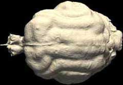 |
Click on the thumbnail to the left for an animation (2.3 mb) of the Felis silvestris catus cranial endocast rotating around the y-axis. |
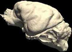 |
Click on the thumbnail to the left for an animation (2.6 mb) of the Felis silvestris catus cranial endocast rotating around the x-axis. |
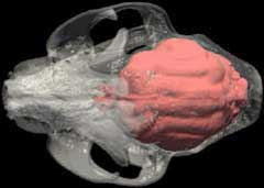 |
Click on the thumbnail to the left for an animation (3.3 mb), rotating around the x-axis, of the Felis silvestris catus cranial endocast highlighted in red within the skull, which is rendered semi-transparent. |
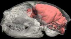 |
Click on the thumbnail to the left for an animation (2.8 mb), rotating around the y-axis, of the Felis silvestris catus cranial endocast highlighted in red within the skull, which is rendered semi-transparent. |
For more endocast imagery, please visit the gray wolf and bottlenose dolphin pages under their respective "Additional Imagery" tabs.
Additional Imagery
 |
|


 )


