|
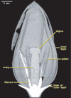
Three-dimensional cutaway reconstruction of the flower of Tulipa sp.. Although high-resolution X-ray CT detects little contrast beyond that between void spaces and tissue, it provides an exact 3D image of the gross morphology of the flower. |

About the Species
This specimen of a tulip, shown below, was obtained from a commercial source in Austin, Texas and was scanned for Dr. Wolfgang Stuppy of the Royal Botanic Gardens, Kew, UK. Funding for scanning was provided by a National Science Foundation Digital Libraries Initiative grant to Dr. Timothy Rowe of the Department of Geological Sciences, The University of Texas at Austin.

About this Specimen
This specimen was scanned by Matthew Colbert on 03 April 2002 along its long axis for a total of 469 512x512 pixel slices. Each slice is 0.0627 mm thick, with an interslice spacing of 0.0627 mm and a field of reconstruction of 30 mm. The outer petals opened during the scan, and therefore were digitally removed prior to rendering.
About the
Scan
Literature
Stuppy, W. H., Maisano, J. A., Colbert, M. W., Rudall, P. J., and T. B. Rowe. 2003. Three-dimensional analysis of plant structure using high-resolution X-ray computed tomography. Trends in Plant Science 8:2-6.
Links
Tulipa on botany.com
tulips on the New York Botanical website
Literature
& Links
None available.
Additional
Imagery
|


 )


)