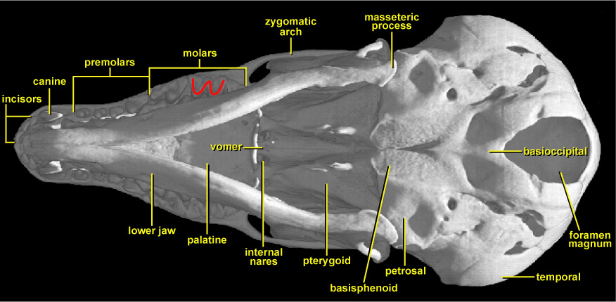
In this view of the skull of Talpa, the two lower jaws obscure much of the secondary palate making up the roof of the mouth. The blade-like nature of the canine teeth at the front of the upper jaws can be appreciated. Between these teeth in the palate lie the palatal shelves of the premaxillae. The molar teeth at the back of the jaws can be seen in crown (occlusal) view. Their cusps are arranged in a distinct W-pattern (indicated in red); this is the dilambdodont dental pattern. This gets its name (literally meaning two lambdas) from the Greek letter for L, lambda λ. When two lambdas are placed together, but upside-down – λλ – they form this arrangement. The dilambdodont pattern of molar tooth cusps is seen in many Mesozoic mammalian fossils and in Talpa represents the retention of a primitive mammalian character. Functionally, the dilambdodont pattern is excellent for the puncture-crushing of tough beetles; the four ridges of the W shape shear past the opposing cusps of the lower molars in a slicing action.
At the back of the secondary palate, between the two halves of the lower jaw can be seen the flat, broad palatine bones on either side of the midline. Immediately behind them are the two internal nasal openings (internal nares or choanae). Separating these two openings is the thin vomer, while on either side of this and further towards the rear of the skull base are the two pterygoid bones. These contact the side wall of the braincase. In the midline of the base of the skull, posterior to the vomer and contacting its rearward tip, is the basisphenoid. On either side of the basisphenoid and close to the jaw joints are the petrosal bones. These are actually separate components of the temporal bones. At the back of the skull on its underside is the very large hole (foramen magnum) for passage of the spinal cord. Between its anterior midline and the back of the basisphenoid lies the basiocciptal. It is partly fused with the basisphenoid and also contacts the petrosal bones. In coronal CT slices of this skull (especially the dynamic cutaways), the scroll-like nasal turbinate bones of the muzzle can be seen to extend far back into the braincase region. In addition, some of the bones such as the frontals, alisphenoids, pterygoids, parietals and petrosals, when seen in coronal section, show similar scroll-like structures. These structures represent pneumatization (air sacs) of these bones; they are also trabeculated (contain small bony rods). This characteristic appearance is well shown in the coronal CT slices.