Coming soon!

About the Species
This specimen (FMNH 45024) was made available to The University of Texas High-Resolution X-ray CT Facility for scanning by Dr. Eric Hilton of the Virginia Institute of Marine Science. Funding for image processing was provided by an National Science Foundation Digital Libraries Initiative grant to Dr. Timothy Rowe of The University of Texas at Austin.
About this Specimen
This specimen was scanned by Matthew Colbert on 13 April 2005 along the coronal axis for a total of 960 slices. Each 1024x1024 pixel slice is 0.1045 mm thick, with an interslice spacing of 0.1045 mm and a field of reconstruction of 49.1 mm.
About the
Scan
Literature
& Links
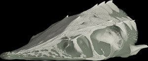
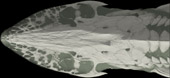
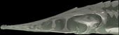
Cutaway animations along the three orthogonal axes - with soft-tissue rendered:
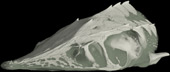
Front page image.
|  |
Additional Imagery
 |


 )


)