Clifford, A. B., and L. M. Witmer. 2004. Case studies in novel narial anatomy: 3. Structure and function of the nasal cavity of saiga (Artiodactyla: Bovidae: Saiga tatarica. Journal of Zoology 264:217-230.
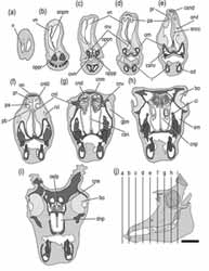
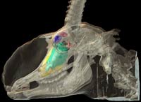
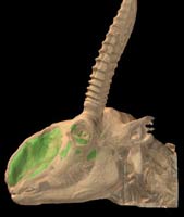
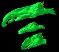
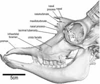
Much of the narial anatomy of the enigmatic antelope Saiga tatarica has been described by previous workers. However, the anatomy of the nasal cavity and the causally-associated osteological correlates of proboscis structure require closer attention, because these data are integral for both a more comprehensive understanding of saiga functional morphology and more robust reconstructions of proboscis structure in fossil taxa. Saiga and outgroup specimens were subjected to X-ray computed tomographic (CT) imaging, gross dissection, and skeletonization. The nasal cavity of saiga is characterised by an enlarged nasal vestibule and basal conchal fold. Many structures (e.g., turbinates, lateral cartilages, mucosal folds, nasolacrimal duct) are retracted caudally to a small area in the caudodorsal part of the nasal cavity proper. The enlarged vestibule is associated laterally and ventrally with paired sacs.
 The nasal septum is largely membranous and contains a large patch of cavernous tissue that serves as a dynamic baffle modifying the flow of inspired air. Bones comprising the narial margin have modified attachment sites for buccinator group muscles and the reduced lateral cartilages. The premaxilla is greatly modified by the enlarged musculature associated with nasolabial fusion. Maintenance of the topological relationships of narial structures compared to bovid outgroups has resulted in a nasal cavity with much larger area for seromucous glands of the vestibule as well as narial musculature capable of controlling the aperture of the nasal cavity. Maxillolabial muscles and the lateralis nasi act together to both
 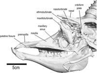 compress the nasal cavity and control the dilation of the nostrils such that air flow through the cavity is highly modified relative to bovid outgroups. The lateral vestibular recess is an outpocketing of the nasal vestibule that produces supplementary seromucous secretions and appears to have no homologue among outgroups. The enlarged nasal vestibule, lateral vestibular recess, repositioned basal fold, and septal cavernous mass are regarded as a coordinated adaptation to dusty habitats, such that nasal air flow can be dynamically regulated allowing for collection of inspired particulates in the vestibule and thus cleansing of air destined for the lungs.
Saiga (Saiga tatarica) are a relatively little studied but morphologically disparate group of antelopes. Bovidae (e.g., antelopes, cattle) have undergone a dramatic radiation in the past 18 million years, now encompassing 135 species (Vrba & Schaller, 2000). Previously of varying affiliation (e.g., Caprinae in Nowak, 1999), saiga are now regarded as members of Antilopinae within Bovidae (Vrba & Schaller, 2000). Saiga have been difficult to place phylogenetically because they are bizarrely apomorphic, particularly in the head and skull as a result of their evolution of unique narial structures. Like many other antelopes, only males possess horns, but their most distinguishing characteristic, a conspicuous proboscis, is possessed by both males and females. The inflated proboscis of saiga provides a case study in the evolution of novel narial anatomy within an otherwise morphologically and phylogenetically well resolved clade.
Saiga are a relatively young species, first occurring in middle Pleistocene deposits approximately 1.0 million years ago, although some authors (Heptner, Nasimovich & Bannikow, 1988) speculated that this genus may occur as far back as the late Pliocene. Only one other species of Saiga is currently recognised (S. prisca), although the Mongolian population previously received species rank (Heptner et al., 1988). Generally, fossil saiga are virtually identical to Recent forms, differing mostly in geographical range (Sokolov, 1974). Occurring now only in open, dusty, arid grasslands of central Asia, their range once extended from the British Isles to eastern Alaska (Frick, 1937).

Literature
Arnautovic, I., and O. Abdalla. 1969. Unusual blind sac on the face of the one-humped camel. Acta Anatomica 73:272-277.
Bannikov, A. G., L. V. Zhirnov, L. S. Lebedeva, and A. A. Fandeev. 1961. Biology of Saiga. Selskokchozyajstvennaya Literature Publications, Moscow.
Blatt, C. M., R. C. Taylor, and M. B. Habal. 1972. Thermal panting in dogs: the lateral nasal gland, a source of water for evaporative cooling. Science 177(4051):804-805.
Boas, J. E. V., and S. Paulli. 1908. The elephant's head: studies in the comparative anatomy of the organs of the head of the Indian elephant and other mammals. Part I. Gustav Fisher, Copenhagen.
Clifford, A. B., and L. M. Witmer. 2001. The narial anatomy of hooded seals (Cystophora cristata) with respect to other Carnivora. Annual Meeting of the Society of Integrative and Comparative Biology, Chicago, Illinois. American Zoologist 40(6):976.
Clifford, A. B., and L. M. Witmer. 2002a. Proboscis evolution in Mammalia: preliminary studies. Annual Meeting of the Society of Integrative and Comparative Biology, Anaheim, California. American Zoologist 41(6):153.
Clifford, A. B., and L. M. Witmer. 2002b. Not all noses are hoses: an appraisal of proboscis evolution in mammals. Annual Meeting of the Society of Vertebrate Paleontology, Norman, Oklahoma. Journal of Vertebrate Paleontology 22(Suppl. to 3):66A.
Clifford, A. B., L. M. Witmer. 2003. Nasal structures in moose (Cervidae: Alces). Annual Meeting of the Society of Integrative and Comparative Biology, Toronto, Ontario, Canada. Integrative and Comparative Biology 42(6):137-138.
Frey, R., and R. R. Hofmann. 1995. Der Kopf der Saiga-Antilope (Saiga tatarica tatarica Linnaeus 1766, Mammalia: Bovidae)---Ausgewählte funktionsmorphologische Aspekte. 1. Die Speicheldrüsen, die Mandibula und die Zunge. Zoologische Beiträge 36(2):169-198.
Frey, R., and R. R. Hofmann. 1997. Skull, proboscis musculature and preorbital gland in the saiga antelope and Guenther's dikdik (Mammalia, Artiodactyla, Bovidae). Zoologischer Anzeiger 235:183-199.
Frick, C. 1937. Horned ruminants of North America. Bulletin of the American Museum of Natural History 69.
Getty, R. 1975. Sisson and Grossman's The Anatomy of the Domestic Animals. I: General, equine, ruminant. (5th edn). W. B. Saunders Company, Philadelphia
Hassanin, A., and E. J. P. Douzery. 2003. Molecular and morphological phylogenies of Ruminantia and the alternative position of the Moschidae. Systematic Biology 52(2):206-228.
Heptner, G. G., A. A. Nasimovich, and A. G. Bannikow. 1988. Mammals of the Soviet Union. I: Artiodactyla and Perissodactyla. Smithsonian Institution Libraries and the National Science Foundation, Washington, D. C.
Jacobi, A. 1921. Die Rüsselbildung bei Säugetieren der Gegenwart und Vorzeit. Jenaische Zeitschrift Naturwissenschaften 50:199-218.
Lodyshenskaya, W. I. 1952. Morphology of the upper respiratory pathways of the saiga antilope (Saiga tatarica). Uchenye Zapiski Karelo-Finskogo Universiteta, Petrazavodsk 4:17-41.
Milner-Gulland, E. J., O. M. Bukreeva, T. Coulson, A. A. Lushchekina, M. V. Kholodova, A. B. Bekenov, and I. A. Grachev. 2003. Reproductive collapse in saiga antelope harems. Nature 422:135.
Murie, J. 1870. On the saiga antelope, Saiga tatarica (Pall.). Proceedings of the Zoological Society, London 1870:451-503.
Nickel, R., Schummer, A., Seiferle & Sack, W. O. (1973). The Viscera of the Domestic Mammals. Siller, W. G. & Stokoe, W. M. (trans.). Verlag Paul Parey, Berlin.<\p>
Nickel, R., A. Schummer, E. Seiferle, J. Frewein, H. Wilkens, and K.-H. Wille. 1986. The Anatomy of the Domestic Animals. I: The Locomotor System of the Domestic Mammals. Siller, W. G. & Stokoe, W. M. (trans.). Verlag Paul Parey, Berlin.
Nomina Anatomica Veterinaria (1994). Ithaca: World Assoc. Vet. Anat.
Nowak, R. M. 1999. Walker's Mammals of the World. (6th edn.). The Johns Hopkins University Press, Baltimore.
Paulli, S. 1900. Über die Pneumaticität des Schädels bei den Säugerthieren. Eine morphologische Studie. II. Über die Morphologie des Siebbeins und die der Pneumaticität bei den Ungulaten und Probosciden. Gegenbaurs Morphologisches Jahrbuch 28:179-251.
Schaller, O. 1992. Illustrated Veterinary Anatomical Nomenclature. Ferdinand Enke Verlag, Stuttgart.
Schmidt-Nielsen, K., F. R. Hainsworth, and D. E. Murrish. 1970. Counter-current heat exchange in the respiratory passages: effect on water and heat balance. Respiratory Physiology 9:263-276.
Sokolov, V. E. 1974. Saiga tatarica. Mammalian Species 38:1-4.
Vrba, E. S., and G. B. Schaller. 2000. Phylogeny of Bovidae based on behavior, glands, skulls, and postcrania; In E. S. Vrba, and G. B. Schaller (Eds), Antelopes, Deer, and Relatives. Yale University Press, New Haven.
Witmer, L. M. 1995. The Extant Phylogenetic Bracket and the importance of reconstructing soft tissues in fossils; pp. 19-33 In Thomason, J. J. (Ed.), Functional Morphology in Vertebrate Paleontology. Cambridge Univ. Press, New York.
Witmer, L. M. 2001a. A nose for all reasons. Natural History 110:64-71.
Witmer, L. M. 2001b. Nostril position in dinosaurs and other vertebrates and its significance for nasal function. Science 293:850-853.
Witmer, L. M., S. D. Sampson, and N. Solounias. 1999. The proboscis of tapirs (Mammalia: Perissodactyla): A case study in novel narial anatomy. Journal of Zoology 249:249-267.


 )