Coming soon!

About the Species
This specimen has no data associated with it. It was made available to the University of Texas High-Resolution X-ray CT Facility for scanning by Dr. Erik Seiffert of Stony Brook University. Funding for scanning was provided by a National Science Foundation grant (BCS-0114856) to Dr. Elwyn Simons of Duke University. Funding for image processing was provided by Dr. Timothy Rowe of The University of Texas at Austin.
About this Specimen
The specimen was scanned by Richard Ketcham on 4 February 2003 along the coronal axis for a total of 957 slices. Each 1024x1024 pixel slice is 0.07896 mm thick, with an interslice spacing of 0.07896 mm and a field of reconstruction of 70 mm.
About the
Scan
Literature
& Links
All endocast image processing by Dr. Ted Macrini, St. Mary's University.
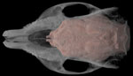 |
Click on the thumbnail to the left for a pitch animation (2.0 mb) of the Procavia cranial endocast highlighted in red within the skull, which is rendered semi-transparent. |
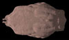 |
Click on the thumbnail to the left for a pitch animation (1.7 mb) of the isolated Procavia cranial endocast. |
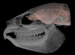 |
Click on the thumbnail to the left for a roll animation (2.6 mb) of the Procavia cranial endocast highlighted in red within the skull, which is rendered semi-transparent. |
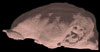 |
Click on the thumbnail to the left for a roll animation (1.8 mb) of the isolated Procavia cranial endocast. |
Additional Imagery
 |


 )


