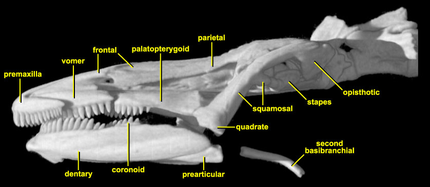
Lateral view of the skull of Necturus maculosus. The lateral view shows the wide anterior premaxilla with its dentition. Behind this, you can see the tooth rows of the vomer, and then the palatopterygoid. Posteriorly, the palatopterygoid abuts the quadrate, itself sheathed laterally by the long, angled squamosal. In this view, you can just see that the sidewall of the skull is solid, but this is most clearly seen (and explained) in the cross-sectional views. Posteriorly, at about its midpoint, the squamosal gives off a short robust process that meets the stylus of the stapes (use the Skeleton Only roll movie to see this more clearly). The footplate of the stapes lies in the large fenestra ovalis, framed by the prootic anteriorly, opisthotic posteriorly, and parasphenoid ventrally.
In this view, the lower jaw is mainly the dentary, although a small part of the prearticular can be seen at the posteroventral edge. The tooth row is a little misleading. Most of the teeth are on the dentary, but the last six teeth are on a separate coronoid bone lying on the medial side of the dentary. Below the skull, and posterior to the jaw, the isolated rod-like element is the second basibranchial of the hyoid apparatus (Walker, 1970, illustrates the hyoid apparatus in its entirety; only the mineralized portion is visible in the CT imagery).