Coming soon!

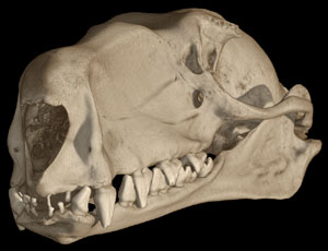
About the Species
This specimen, a male, was made available to The University of Texas High-Resolution X-ray CT Facility for scanning courtesy of Dr. Nancy Simmons of the American Museum of Natural History. Funding for scanning was provided by a National Science Foundation grant (DEB-9873663) to Dr. Simmons, and funding for scanning and image processing was provided by a National Science Foundation Digital Libraries Initiative grant to Dr. Timothy Rowe of the Department of Geological Sciences, The University of Texas at Austin.
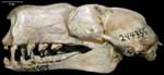
About this Specimen
This specimen was scanned by Matthew Colbert on 28 April 2005 along the coronal axis for a total of 1625 slices. Each 1024x1024 pixel slice is 0.04465 mm thick, with an interslice spacing of 0.04465 mm and a field of reconstruction of 42.5 mm.
About the
Scan
Literature
& Links
Front page image.
|  |
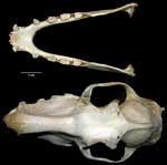
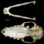
Additional
Imagery
|


 )


)