Dromaius novaehollandiae, the emu, is a paleognath bird. This is a group of large, flightless birds that also includes the extinct elephant bird and moa and the extant ostrich, rhea, kiwi, and tinamou. Emus are indigenous to Australia, and have recently been raised in captivity for their eggs, meat, and skin. Emus usually weigh between 50-55 kg, and have an average height of 1.75m, making them the second largest living bird. Like ostriches, emus are very fast, capable of top speeds around 50 km/hr; they are also strong swimmers. The emus diet generally consists of seeds, fruits, and young shoots, but they will eat insects and small vertebrates when available. (Information obtained from the University of Michigan Museum of Zoology page - see links).
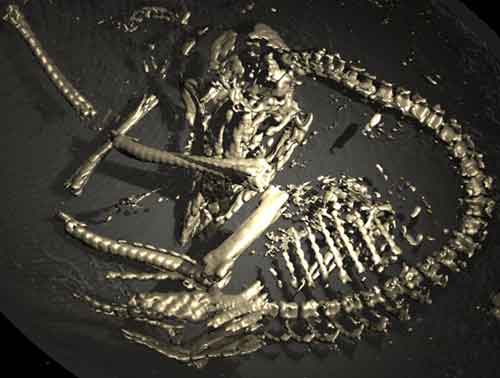 | VGStudio Max visualization produced by Christof Reinhart of Volume Graphics GmbH. |

About the Species
This specimen was highlighted in the February 23, 2001 issue of Science magazine.
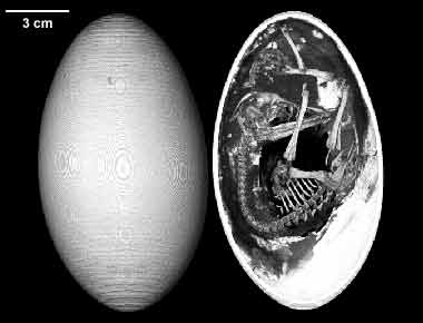
The sample for this study consists of a frozen emu egg with a late term embryo inside. The specimen was donated to the Vertebrate Paleontology Laboratory of the Texas Memorial Museum by Gerald Grellet-Tinner. Funding for its scanning at The University of Texas High-Resolution X-ray CT Facility was provided by Dr. Timothy Rowe.
About this Specimen
The Dromaius egg was scanned by Richard Ketcham and Cambria Denison in September of 1997 along its long axis for a total of 279 slices, each slice 0.5 mm thick, with an interslice spacing of 0.5 mm. The dataset displayed was reduced for optimal Web delivery from the original, much higher-resolution CT data. A sample slice, shown below from the original dataset, is through the skull of the embryo and illustrates the detail discernible in the unreduced CT data.
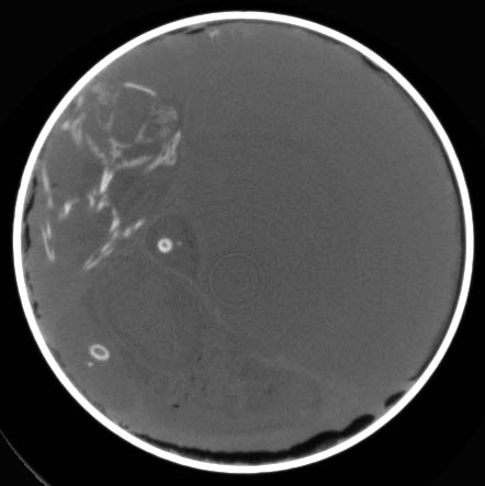 |
| Sample slice #108 through the skull of the embryo inside the egg. |
About the
Scan
Literature
Baumel, J. J. (ed.) 1993. Handbook of Avian Anatomy: Nomina Anatomica Avium. Second edition. Nuthall Ornithological Club Publication, Cambridge, Massachusetts. 779 p.
Carpenter, K., Hirsch, K. F., and J. R. Horner (eds.). 1994. Dinosaur Eggs and Babies. Cambridge University Press, New York. 372 p.
Carroll, R. L. 1988. Vertebrate Paleontology and Evolution. Freeman, New York. 698 p.
Chiappe, L. M. 1996. Early avian evolution in the Southern Hemisphere: The fossil record of birds in the Mesozoic of Gondwana. Memoirs of the Queensland Museum 39:533-555.
Chiappe, L. M. 1995. The first 85 million years of avian evolution. Nature 378:349-355.
Cracraft, J. 1973. Phylogeny and evolution of the ratite birds. Ibis: 116.
Gill, F. B. 1995. Ornithology, second edition. W. H. Freeman, New York. 763 p.
Links
Dromaius novaehollandiae on The Animal Diversity Web (The University of Michigan Museum of Zoology)
Dromaius novaehollandiae on the Oakland Zoo website
Dromaius novaehollandiae on the Unique Australian Animals website
Dromaius novaehollandiae from the Virginia Emu Association
Literature
& Links
None available.
Additional
Imagery
|