|
|
 Expert annotations for this species! See below. Expert annotations for this species! See below.
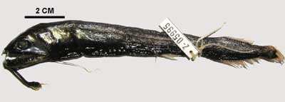
Chirostomias pliopterus is a relatively primitive member of the Stomiidae, a group called scaleless dragonfishes. These fishes are primarily mesopelagic, meaning that most inhabit open ocean waters about 1000 to 3000 meters deep, although some come to the surface at night to feed and others stay in waters as deep as 5000 meters. Plants do not live at these depths, so all dragonfishes are predators on other fishes and deep-water invertebrates.
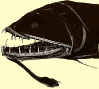 |
Head of Chirostomias pliopterus showing barbel attached to lower jaw |
|
Among the specializations of dragonfishes are very large hinged teeth, light organs on a barbel (a fleshy extension from the lower jaw) and/or on the pectoral fins, and in some species modifications that resemble a spring on the anterior portions of the backbone. In addition, most dragonfishes have small light organs, some forming lines, along the body. The feeding apparatus of these animals is well developed, but the rest of the body is less so, and reduced skeletons and musculature are specializations associated with the low energy budget of the mesopelagic environment.
Chirostomias pliopterus is especially noteworthy because of the pectoral fin is highly modified to carry an elaborate light organ. It is otherwise one of the most morphologically primitive members of the group. It occurs in the Atlantic Ocean from approximately 20°N to 45°N. Most individuals have been captured above 1300 meters. There is very little known about this species, as few individuals have been captured, and those few have been little studied.
|
The skeleton of Chirostomias is lightly mineralized, with the exception of the jaws and other portions of the feeding apparatus, which are the most distinctive features of the scan. These bones include the premaxilla and maxilla of the upper jaws and the dentary and anguloarticular of the lower jaw. In addition, the major bones that support or reinforce the jaws, including the hyomandibula and preopercle, are easily visible. The pectoral girdle bones, which are used both in locomotion and feeding, are also well developed, including the posttemporal and cleithrum. |
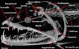 |
Head skeleton of Chirostomias pliopterus. |
|
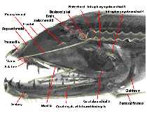 |
Head of Chirostomias pliopterus, sagittal cutaway view, with portions of the soft tissues visible. |
|
The jaw teeth of Chirostomias are large and recurved. Some are tightly bound to the jaw bones, while others are hinged, allowing them to tilt back when prey are pulled backwards towards the gullet. Like most other fishes, Chirostomias has a second set of jaws deep in the oral cavity on certain bones of the branchial basket, and these are clearly visible in the scan. The branchial basket carries the gills as well as teeth, and serves several important functions in the life of the individual. Some of the bones of this basket can be seen in the scan, mostly as small fragments distributed in the oral cavity.
|
Most of the braincase is cartilage and not visible in the scan; the bones surrounding the brain are very thin and lightly mineralized so are not clearly visible or do not appear at all. The vertebral column is clearly visible in the specimen. Perhaps less obvious is the fact that the anterior centra are smaller and slightly less mineralized than those more posterior. In most stomiids, the anterior centra are either very lightly mineralized or not present at all, leaving the prominent notochord as the major support between the head and postcranial body. Also visible are the small, multi-branched intermuscular bones behind the head and along the vertebral column. These bones lie in the muscles of the body and serve as anchors for them.
The pectoral fins are visible, as are some of the small bones, the pectoral radials, which support and connect them with the pectoral girdle. Chirostomias has unique pectoral radials, associated with the specialized morphology of the fins.
Other distinctive features of the scan are the eye lenses, which are composed of dense proteins, and the otoliths (ear stones), which are densely mineralized structures used in hearing and balance.

About the Species
This specimen (UMMZ 227717) was collected by K.E. Hartel on the RV Oceanus in the North Atlantic Ocean at a depth of 800m. It was made available to the University of Texas High-Resolution X-ray CT Facility for scanning by Dr. William Fink of the University of Michigan. Funding for scanning and image processing was provided by a National Science Foundation Digital Libraries Initiative grant to Dr. Timothy Rowe of the Department of Geological Sciences of The University of Texas at Austin.
About this Specimen
The specimen was scanned by Matthew Colbert on 9 June 2003 along the coronal axis for a total of 945 1024x1024 pixel slices. Each slice is 0.0348 mm thick, with an interslice spacing of 0.0348 mm and a field of reconstruction of 32.7 mm.
About the
Scan
Literature
Fink, W.L. 1985. A phylogenetic analysis of the family Stomiidae (Teleostei, Stomiiformes). Miscellaneous Publications of the Museum of Zoology, University of Michigan, 171:127 pp., 70 figs.
Fink, W.L. 1998. Dragonfishes and their allies. In: Encyclopedia of Animals: Fishes, J.R. Paxton (Ed.). Weldon Owen, Australia. (Second Edition; Encyclopedia of Fishes, J.R. Paxton and W.N. Eschmeyer, eds.), 119-122). Academic Press, San Diego, California, 119-122.
Morrow, J.E. and R.H. Gibbs. 1964. Family Melanostomiatidae. In: Fishes of the Western North Atlantic. Part 4. Sears Foundation for Marine Research. Pp. 351- 511.
Literature
& Links
None available.
Additional
Imagery
|
|


 )